What does COVID look like on a chest xray?
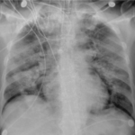
(RSNA) Case example of diffuse non-specific airspace opacities in both lungs in an intubated patient with confirmed COVID diagnosis on day 6...
Coronal Chest CT: COVID demonstrating Nodular and Mass-like Airspace Opacities
(RSNA) - CT Chest coronal view demonstrates multifocal nodular and mass-like airspace opacities in both lungs. Source: Song et al, Radiology...
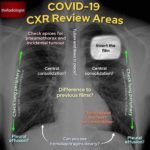
How to read a COVID-19 Chest X-Ray – visual guide
How to read a COVID-19 Chest X-Ray - visual guide
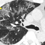
Radiologic Sign of the Day (CT) for COVID: Crazy Paving
On cross sectional CT imaging of the lung, sometimes, you might see what is known as thickened interlobular and intralobular lines (opacities)...
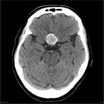
Case of the Day: Brain Mass on CT
What is your diagnosis? A. Craniopharyngioma or B. Aneurysm
Answer - Click here
Chest X-Ray Basics: PA vs. AP
Which one is AP and which one is PA? The answer is... below.
On the PA view,...
Lipoid Proteinosis (hyalinosis cutis et mucosae or Urbach-Wiethe disease)
Lipoid Proteinosis
Waxy, thickened, yellowish skin with multiple permanent, poxlike atrophic scarring on the forehead. Source: Dr....
Cutaneous findings in systemic sarcoidosis
Cutaneous findings in systemic sarcoidosis
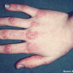
Dermatomyositis
Dermatomyositis
Tinea Corporis
Tinea Corporis
Annular erythematous scaling patch with an active border composed of papules, vesicles and crusts.

