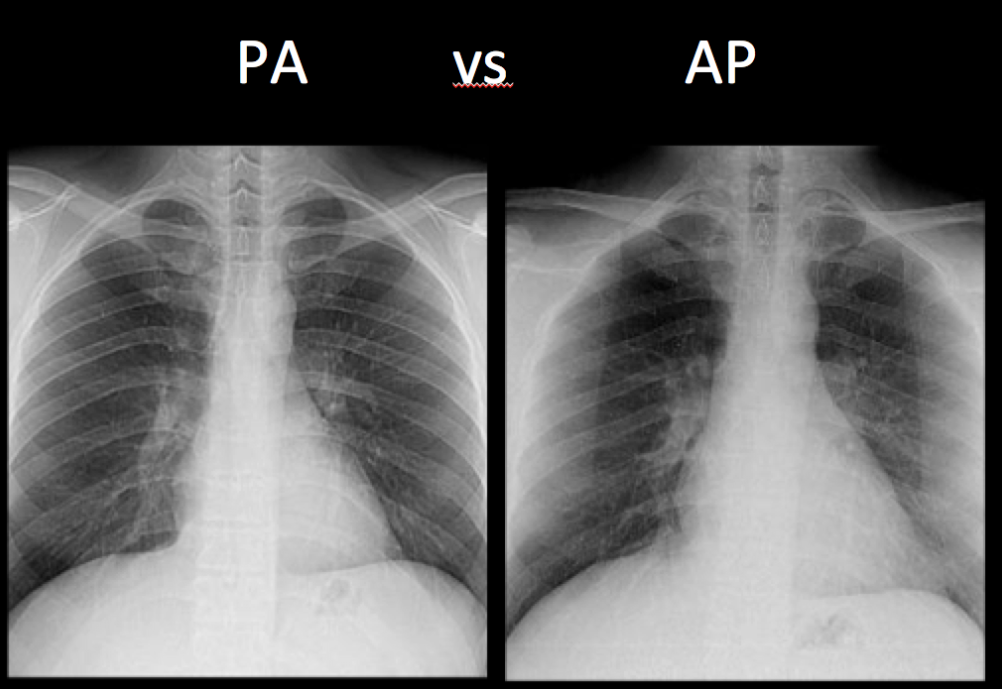
On the PA view, the cardiac borders are smaller and more defined. Given the way the x-ray beam works, the heart appears smaller and with sharper borders on the PA view. The reason is that the patient’s chest (anterior) is against the x-ray film with the beam entering from posterior (P) to anterior (A) – hence the term “PA.” Similarly, the AP view is when the beam enters from front to back with the x-ray film at the back of the patient – therefore, the heart is magnified and the margins are minimally less sharp.


