Social Rounds Feature from social media! Social Rounds is where we feature interesting and educational cases on social media. Discover new cases and profiles you should follow too!
This case came across the wires on Instagram – Follow @globaldermie who recently posted this most interesting case!
Diagnosis: Tuberous Sclerosis
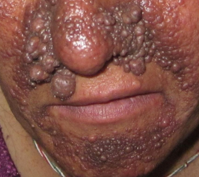
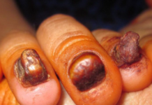
Case description: 40 yo female. Developing country. Diagnosis: tuberous sclerosis (TS). Intellectual disability. Photos: angiofibromas and ungual fibromas. CT scan: multiple calcified subependymal nodules along margins of lateral ventricles. Autosomal dominant inherited genetic disorder characterized by hamartoma formation in multiple organs caused by mutations in genes TSC1 encoding hamartin and TSC2 encoding tuberin (which form a complex involved with PI3K signaling pathway, regulates cell growth/proliferation). 2/3 occur due to spontaneous mutation. TS frequently affects heart, kidneys, nervous system, and skin. Diagnosis may be difficult, subtle. Manifestations include facial angiofibromas, developmental delay, and seizures (Vogt’s triad seen in 33%). Waxy papules (angiofibromas) develop on cheeks, nose, forehead. Hypomelanotic “ash leaf” macules occur on extremities and trunk. Thick plaques with orange-peel-like consistency occur on the trunk, especially lumbosacral area, called shagreen patches and correspond to collagenomas. Periungual/ungual and gingival fibromas occur with less frequency. Wood’s lamp may accentuate ash-leaf macules and help diagnosis, often present at birth or shortly after. Diagnosis based on major and minor criteria. Definite TS: 2 major features, or 1 major and >=2 minor features. Possible TS: 1 major or >=2 minor features. Major features: angiofibromas (>=3) or fibrous cephalic plaque, hypomelanotic macules (>=3, at least 5mm), shagreen patch, cardiac rhabdomyoma, cortical dysplasias, subependymal giant cell astrocytoma, subependymal nodule, ungual fibromas (>=2), multiple retinal nodular hamartomas, angiomyolipomas (>=2), lymphangiomyomatosis. Minor features: intraoral fibromas, multiple renal cysts, “confetti” skin lesions, dental enamel pits (>=3), retinal achromic patch, and nonrenal hamartoma. Facial angiofibromas can be treated with cryotherapy, surgical, or laser therapy. Topical mTOR inhibitor, rapamycin is also effective, and can also be used to treat ungual fibromas. #mysterydiagnosis #globalderm #globaldermatology #tuberoussclerosis #genoderm #dermboards #tbcd #dermed
Source: @globaldermie


